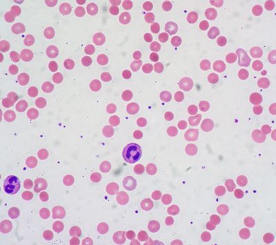
Hemolysis often presents with chronic or acute anemia, jaundice, or radiculopathy.

Hemolytic anemia is usually caused by increased serum albumin concentration. The diagnosis of hemolytic anemia is made by elevated haptoglobin level, increased unconjugated bile, jaundice, and decreased haemoglobin S-100 in the blood. In patients with jaundice, increased bilirubin, increased serum albumin, or low hemoglobin can cause hemolytic anemia. In patients with radiculopathy, reduced levels of both soluble and insoluble collagens can produce this condition.
Hemolytic anemia can be a result of the presence of the hemolytic enzymes leukotriene-conjugating enzyme (LTCE) in the bloodstream. When there is an increase in the production of LTCE, it can lead to the increased excretion of heme, resulting in increased serum albumin concentration and elevated levels of circulating ferritin. Increased serum albumin concentration and high levels of circulating ferritin can also contribute to hemolytic anemia.
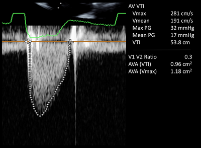
Normal hemoglobin content can vary widely from one person to another. Therefore, the determination of the proper hemoglobin levels in patients suffering from hemolytic anemia is very difficult, particularly in patients with normal serum albumin concentration. It is usually recommended that patients suffering from hemolytic anemia should have their serum albumin levels measured by means of Doppler ultrasonography.
In addition to measuring the levels of albumin, Doppler ultrasonography can also be used to measure blood pressure and cardiac output. The results of the measurement of these variables can help in the diagnosis of hemolytic anemia and the treatment of hemolytic anemia. The results are usually expressed as mean values or medians. The most accurate values for hemolytic anemia will usually depend on the total number of units of hemoglobin and the percentage of hemoglobin that is present in the red blood cells.
Hemoglobin is the primary protein found in red blood cells.

Hemoglobin is separated from the plasma and transported to other organs when blood is pumped out. There are several types of hemoglobin.
Patients suffering from hemolytic anemia can be diagnosed based on the type of hemoglobin present in their blood, known as the hemoglobin type. There are four types of hemoglobin: A, B, AB, O, G2, and T. A, B, O, and G2 are the four different types of hemoglobin. Blood cells can have either of these four types of hemoglobin present in their membrane, but the most common types are usually A, B, O, G2, and T. Each type of hemoglobin is different in their effects on health and their respective properties.
Because hemoglobin is the substance responsible for the transport of oxygen to tissues, when hemoglobin is not present, the blood cannot carry oxygen and other nutrients to other areas of the body. Anemia develops due to anemia. The red blood cells carry less oxygen to other parts of the body, causing an increased risk of infection, anemia, or even anemia due to decreased blood supply to the brain. Anemia can cause damage to the organs, such as the brain or heart, when the blood supply is not sufficient for adequate amounts of oxygen to pass through the blood vessels, damaging the cell structure and increasing the risk of death.
In patients suffering from hemolysis, measuring the levels of albumin and measuring the levels of hemoglobin may be helpful in diagnosing hemolytic anemia and determining whether treatment may need to be sought to increase levels of these substances. The results of a Doppler ultrasonography test are used to help in the diagnosis and treatment of hemolytic anemia. The results of the test will help in the diagnosis of hemolytic anemia and the treatment of hemolytic anemia when the patient has anemia due to hemoglobin insufficiency and there is an increased risk of infection, anemia due to increased blood supply to the brain, or when there is an increase in the risk of a blood clot forming in the brain.